
Joint surface damage (Chondral defects / osteochondral defects)
What is it?
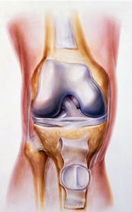
The knee joint is a type of joint known as a synovial joint. A synovial joint is a connection of two bones together through a moving articulation. At the end of each bone there is a joint surface (articular surface) that is lined by a very low friction material called articular cartilage. This cartilage is lubricated by a film of fluid called synovial fluid that is produced by a very thin fragile lining called the synovium. The synovium is supported by a much stronger fibrous structure that contains the whole joint and is called the joint capsule.
Any part of a joint can be injured and a common injury to the knee is a localised loss of articular cartilage (chondral defect) sometimes combined with an injury to the underlying supporting bone (osteochondral defect).
When does it occur?
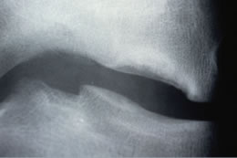
Osteochondral defects can occur spontaneously often in the growing or young adult skeleton (sometimes known as osteochondritis dissecans / OCD) or can occur as a result of an injury or trauma to the knee. The trauma may be of a very minor nature particularly in the more mature knee and may be associated with a more generalised wear and tear process within the joint.
What are the symptoms?
Most people will describe an insidious onset of intermittent pain, swelling and catching of the joint. There may be a feeling of instability or the knee may fail to fully straighten.
Symptoms will probably be worsened by activity and eased by rest.
What is the treatment?
There are a large number of variables that influence the approach to treatment. Some people will merely need an exercise regime and modification of activity, whereas others may need surgical intervention. The timing of surgery will also vary significantly. There are many operations used to treat the various types of this condition.
Arthroscopy (keyhole surgery)
If there is a partial thickness injury to the articular cartilage and mechanical symptoms of catching and intermittent acute pain persist, it may be appropriate to carry out an arthroscopy and smooth the joint surface to remove loose cartilage (debridement / chondroplasty)
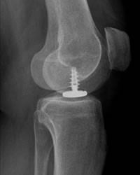
Re-attachment (internal fixation)
Occasionally the damaged area of cartilage and its underlying bone (osteochondral fragment) will remain partially attached or be loose but in one piece within the joint. In this situation, fixation of the fragment back into the defect is, if possible, desirable.
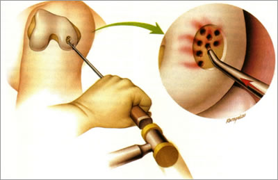
Microfracture
In the presence of a full thickness cartilage defect the aim is to attempt to restore a cartilage covering to the underlying bone. Articular cartilage has no blood supply and therefore has no ability to heal once it is damaged. Microfracture is an operation done with keyhole surgery where multiple small holes are created in the bony base of the defect allowing blood to enter the cavity and initiate a healing response. The post operative treatment is the key to the success of the procedure.
Other operations are also designed to replace, repair or restore damaged articular cartilage.
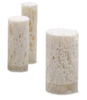
Synthetic plugs
There are a variety of man made implants that can be placed into defects either with keyhole or minimally invasive surgery. These aim again to encourage a healing response and may be designed to disappear over time as the healing tissue forms.
Grafts
In general grafts are pieces of natural tissue taken from another place and implanted into a defect. Grafts can come from yourself (autograft), another person (allograft) or a different animal (xenograft).
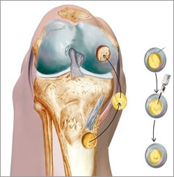
OATS (osteoarticular transfer system)
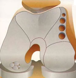
This is an autograft technique where cylinders of articular cartilage and its underlying bone are taken from less crucial parts of the knee joint surface and replaced as multiple plugs into holes drilled into the floor of the defect. The result is rather like that of cobble stones.
A similar procedure can be carried out with slightly larger allograft plugs giving a smoother effect although supply, matching joint curvature and sterilisation become an issue.
Autologous chondrocyte implantation (ACI / MACI)
There is a lot of research into ways of restoring normal healthy articular cartilage into joint surface defects. Cartilage can be made either using cells that have already become cartilage cells (chondrocytes) or using cells usually from the blood or bone marrow that have not yet become a specific cell type (stem cells).
Autologous chondrocyte implantation involves two operations. The first is used to take a small amount of healthy cartilage from the knee through keyhole surgery. This tissue is sent to a laboratory where the chondrocytes are grown and multiplied. Once enough cells are present they are re-implanted into the joint surface defect either beneath a membrane (ACI) or within a matrix or scaffold (MACI). The ultimate aim is that healthy articular cartilage is restored. The procedures are evolving continually as new technology becomes available.
Partial surface replacement
An alternative solution to restoration or repair is to replace the lost articular cartilage with a man made bearing. With small defects of the joint surface the entire compartment of the knee should not be replaced. There are new implants coming to the market aimed at replacing small areas of the joint surface. Little at present is known about the outcome of this approach and early results are awaited.
Bristol
Contact us on
0117 980 4037
or
0117 980 4046